V3 - Implantat mit konischer Verbindung
Das V3-Implantat ist eine bahnbrechend innovative und ausgeklügelte Lösung. Es wurde von Zahnärzten für Zahnärzte entwickelt – mit einer Geometrie, die durch Gewebeerhalt und -wachstum für Verfahren mit optimalen ästhetischen Ergebnisse ausgelegt ist.
Die MIS-Lösung mit konischer Verbindung bietet:
- Eine einheitliche Prothetik
- Ein Chirurgie-Set
- Ein Bohrprotokoll
- Zwei spezifische Geometrien der C1- und V3-Implantatsysteme sorgen für optimale Implantatintegration und Knochenwachstum.
Vorteile
- Ästhetik: Ein breites Sortiment an Prothetik-Komponenten für die konische Verbindung von MIS bietet: Kompromisslose Präzision; ein gleichmäßiges konkaves Emergenzprofil für exzellente Ergebnisse hinsichtlich des Weichgewebes; goldenen Farbton zur Unterstützung hochästhetischer Ergebnisse.
- Implantatintegration: Der dreieckige Hals des V3 wurde so konzipiert, dass ein Reservoir für eine Blutansammlung und die Bildung von Blutgerinnseln entstehen kann. Diese Bedingungen sind die Voraussetzung für eine optimale Implantatintegration und das Knochenwachstum.
- Knochenerhalt: Die Lücken an den Seiten des Implantathalses wurden so gestaltet, dass daraus eine offene, kompressionsfreie Zone resultiert. Der krestale Knochenverlust kann durch Abbau der Belastung im Kortikalisknochen minimiert werden.
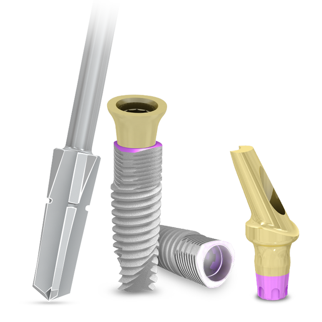
- Maximale Präzision: Jedes V3-Implantat wird im Paket zusammen mit einem sterilen Finalbohrer für einmaligen Gebrauch geliefert; der Bohrer ist für alle Knochentypen geeignet und erhöht die Wahrscheinlichkeit für eine präzisere Passung. Die V3-Eindrehinstrumente sind mit Markierungen versehen, um die Ausrichtung des Implantats bei der Insertion zu vereinfachen.
- Klinischer Erfolg: Die rauhe Oberflächenbeschaffenheit und die Mikromorphologie sämtlicher MIS-Implantate ist das Ergebnis eines Sandstrahl- und Säureätzverfahrens. Diese bewährte Oberflächentechnologie von MIS hat bereits Millionen von Patienten ausgezeichnete Ergebnisse bei der Osseointegration und lang anhaltende klinische Erfolge beschert und beruht auf jahrelanger Forschung und entsprechend belegbaren Daten.
V3 mit B+ Oberfläche
B+ ist ein biologisches Merkmal der MIS-Implantate, das zu einer effektiven Langzeit-Osseointegration führt. Eine monomolekulare Schicht aus Multi-Phosphonaten, die permanent auf der Implantatoberfläche gebunden ist, wird vom Körper als knochenartige Struktur erkannt.
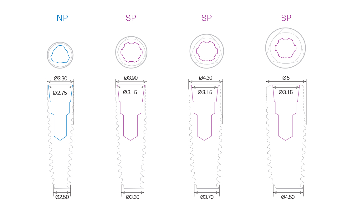
Technische Information


